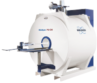
Small Bore 7T Imaging
For preclinical imaging, we have a small-bore 7 T MRI. An introductory applications brochure has been prepared.
The 7T MRI lab houses an actively shielded Bruker BioSpec 70/30 USR 7T system with the Bruker AVANCE III HD architecture and Paravision 6 Software. This state-of-the-art scanner can reach a spatial resolution of 25 microns.

Available RF-probes include a 1H MRI Cryoprobe, and a 1H receive-only 8 x 1 rat head surface array coil.
Gradients available are:- BGA 20 (200 mT/m, ID 200 mm)
- BGA 12 (400 mT/m, ID 120 mm)
- BGA 9S (675mT/m, ID 90 mm)
- BGA 6 (1000 mT/m, ID 60 mm)
(ID = maximum usable internal diameter)
Examples of methods which can be used on the 7T system include:
- 'Conventional' MR images based on T 1, T 2 or proton density, typically used to show anatomical detail;
- Blood flow in arteries or veins (magnetic resonance angiography or MRA);
- Blood perfusion through tissue, giving cerebral blood flow (CBF) and cerebral blood volume (CBV) maps;
- Molecular diffusion of water through tissue such as white matter tracts (tractography and diffusion tensor imaging (DTI));
- Relative degrees of bound and unbound water via magnetization transfer contrast (MTC);
- Tissue movement, such as motion of the heart to yield measures of ejection fraction and myocardial wall motion;
- Tissue temperature and intracellular pH measurements;
- Oxygenation of blood to show areas of brain activated by stimuli - functional MRI (fMRI);
- Changes in blood perfusion through tissues responding to pharmacological intervention (phMRI);
- Cell tracking following the labelling of cells with magnetic nanoparticles.
- Multi-nuclei spectroscopy and imaging is possible.
Access Charges
€1,200 per day
A limited number of discounted slots are available, for pilot or training studies.
Contact
7T MRI contact number: 01 896 1487

